A 65 y.o. nurse with a hx of diabetes presents with arthritis and elevated liver enzymes.
What would you order?

Hint: she notes her skin is darkening slightly on her face.
Our patient had hemochromatosis.
Bronzing of the skin is a classic description of hemochromatosis although this can occur late in the disease and look like a suntan. Bronzing of the skin is caused by a deposition of iron in the sweat glands. Iron overload also causes an increase in free radical activity and end organ damage of the pancreas, liver, kidneys and heart. Diabetes and cirrhosis of the liver are common. Rarely, hypocalcemia may result from infiltration of the parathyroids.
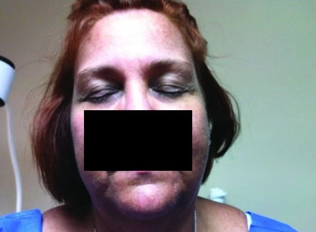
bronzing of the eyelids from hemochomatosis
Hemochromatosis is one of the most common genetic disorders in the United States. The increased iron results from one of two main causes:
Hereditary hemochromatosis is an autosomal recessive disorder associated with overexpression of HRF gene on chromosome six regulating the intestinal absorption of iron.
Secondary hemochromatosis results from repeated transfusions. This can occur in sickle cell disease or transfusions for aplastic anemias.
THE DIAGNOSIS
The diagnosis is difficult early in the course of the disease because the symptoms can be nonspecific e.g. fatigue, diabetes or joint pain. If there is a suspicion, a ferritin will help with the diagnosis. Ferritin is high as well as serum transferrin saturation (TSAT) The transferrin saturation is generally above 45% because the iron-carrying protein is saturated with the excess circulating iron. Total iron binding capacity(TIBC) is low because the binding sites on transferrin are already full.
In addition to those receiving iron, certain populations are at risk. Northern European populations have the highest incidence of the disease. Excessive use of vitamin C in health- conscious people is associated with iron overload because Vit C increases the absorption of iron in the gut. Excessive alcohol use can exacerbate hereditary hemochromatosis since alcohol disrupts iron regulation leading to increased iron absorption and storage in the liver.
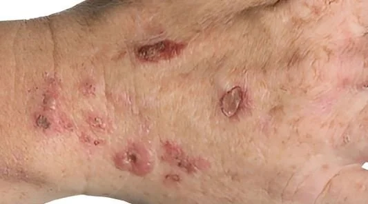
Porphyria cutanea tarda is a blistering rash found in alcoholics and hemochromatosis
Normal total body iron is 2.5 gr. In females and 3.5 gr in males. Because symptoms may be delayed until iron overload is excessive e.g., 10 to 20 grams, it is often not recognized until later in life. In women it is unlikely to present before menopause. In these hereditary forms liver disease is the most common complication and may progress to cirrhosis. Cardiomyopathy with heart failure is the second most common complication. Porphyria cutanea tarda may result since excess iron inhibits the liver enzyme uroporphyrinogen decarboxylase (URO-D) leading to a build up of pophyrins which trigger skin blisters. Classically, arthropathy with chondrocalcinosis of the second and third metacarpals may result.
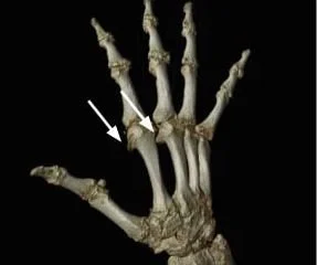
arthropathy of the second and third metacarpals in hemochromatosis
Our patient tried to donate blood. It was discovered that her ferritin was 800. She had a liver biopsy which confirmed the diagnosis showing increased iron in hepatocytes, but did not show cirrhosis. She was initially treated with phlebotomy. She was advised to avoid red meat and excessive amounts of Vit C. She is being followed by her physician.
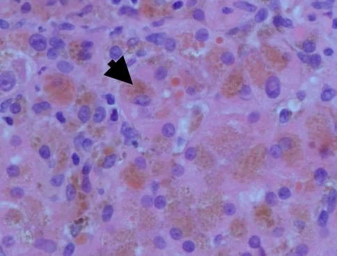
liver biopsy showing iron in the hepatocytes.
Hemochromatosis.org is a digital education and awareness effort provided
by Iron Disorders Institute, a 501 c (3) non-profit agency.
lRichardi D, Brown EM. Physiology and pathophysiology of the calcium-sensing receptor in the kidney. Am J Physiol Renal Physiol. 2010;298:F485–99.
Elgendy I, Omokehinde T. A rare cause of hypocalcemiaAm J Case Rep 2013 Apr 19;14:113-115.
Chacon S, Morrison B, Shasa H. Acquired hemochromatosis with pronounced pigment deposition of the upper eyelids. J Clin Aesthet Dermatol. 2013 Oct ;6(10):44-46.