A 50 y.o. bird watcher states she is having trouble seeing the birds because of black spots in her vision.
When you look in her eyes you see black spots. What is happening?
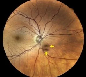
The patient reports the black spots move when she moves her eyes to follow a bird.
Our patient has myodesopia better known as “floaters” on ophthalmoscopy. These are the shadows of tiny clumps of gel in the vitreous humor which appear on the retina. They will appear out of focus because they are actually in front of the retina. From the patient’s perspective floaters are small dark specks or squiggly lines drifting across the field of vision. When patients try to focus on them, they dart away, moving with their eyes. They are most noticeable when looking at a plain background like a blue sky which is why our bird watcher complained that there were two birds in the sky but she could not focus on one.
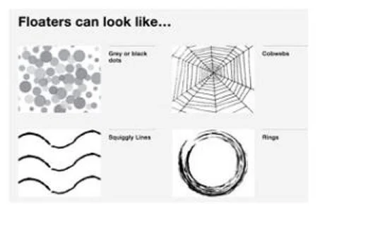
types of floaters
As the vitreous ages it becomes thinner and at some point pulls away from the retina. This is more likely in near-sighted individuals. Flashes occur with traction on the retina and this is known as posterior vitreous detachment. Floaters can be most easily seen by illuminating the retina through the pupil (think; the red reflex) The light beam of a slit lamp and the microscope are placed in a similar axis instead of at 45 degrees. The light passes through the pupil and creates a background glow; floaters appear as shadows in the vitreous.
Lasers have been developed to treat certain types of floaters. This is known as laser vitreolysis (aka LFT). It can be performed in office and does not require anesthesia since the procedure is painless. It works very well for patients who are troubled by the “annoying” symptoms of floaters. Neuroadaptation occurs with most floaters and in three to six months. Your visual system learns to suppress them. They are no longer seen.
For those floaters that do not qualify for laser, and limit vision, surgery is required to replace the vitreous gel. This procedure, known as a pars plana vitrectomy, removes the natural vitreous and replaces it with fluid.
Lasers have been developed to treat certain types of floaters. This is known as laser vitreolysis (aka LFT). It can be performed in office and does not require anesthesia since the procedure is painless. It works very well for patients who are troubled by the “annoying” symptoms of floaters. Neuroadaptation occurs with most floaters and in three to six months. Your visual system learns to suppress them. They are no longer seen.
For those floaters that do not qualify for laser, and limit vision, surgery is required to replace the vitreous gel. This procedure, known as a pars plana vitrectomy, removes the natural vitreous and replaces it with fluid.
FLOATERS MUST BE DIFFERENTIATED FROM RETINAL DETACHMENTS AND AI CAN MAKE THIS MORE DIFFICULT FOR A CLINICIAN
Patients often consult the internet before seeing a physician. A recent patient was described by Bair. She reviewed her symptoms on the internet and AI decided her symptoms of light flashes were compatible with a migraine. The chatbot offered a plausible explanation for her symptoms . It asked leading questions and created a single diagnosis rather than ten possible diagnoses in a differential. The questions AI asked were:
Did the light zigzag across your vision?
Did it last 20 minutes?
Was there a headache afterwards?
When seen by Dr. Bair, a retinal detachment was discovered and she had immediate laser surgery. It took the physician going back to the original symptoms to make the diagnosis. The flashes had occurred at night and the headache was not the predominant symptom.
In a study from MIT and UC Irvine by Loftus, people who recalled an event by a back-and-forth with a generative AI chatbot formed three times as many false memories as a control group. Suggestion creates recollection and anchoring on a previous diagnosis prevents obtaining the correct diagnosis by both the patient and the physician.
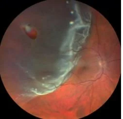
flashes of light are found in both posterior vitreous detachment and retinal detachment
Our patient with floaters had a retinal exam which was normal and in follow up at 3 months her floaters were gone.
https://www.sciencedirect.com/topics/medicine-and-dentistry/vitreous-floaters
Yang X, Shi C, Liu Q. spontaneous remission of vision degrading myodesopsia of posterior vitreous detachment . Graefe’s archive for clinical and Experimental Ophthalmology (2023)261:1571=1477. https://doi.org/10.1007/s00417-022-05948-4.
Serpetopoulos CN, Korakitis RA (1998) An optical explanation ofthe entoptic phenomenon of ‘clouds’ in posterior vitreous detachment. Ophthalmic Physiol Opt 18:446–451 .
Bair H. AI doesn’t just misinform patients, it gives them a plausible story. June 30, 2026.
Yorston D, donachie P, Laidlaw D, et al. Predicting post-surgery change in visual acuity after successful repair of macula-off retinal detachment: findings from a large prospetive UK cohort study. Eye. 2025. 39, 2298-2306.
Pataranutaporn P, Archlwaranguprok C, Chan S, Luftus E, et al. Synthetic human memories: SI-edited images and videos can implant false memories and distort recollection In Proceedings of the 2025 CHI Conference on Human Factors in Computing