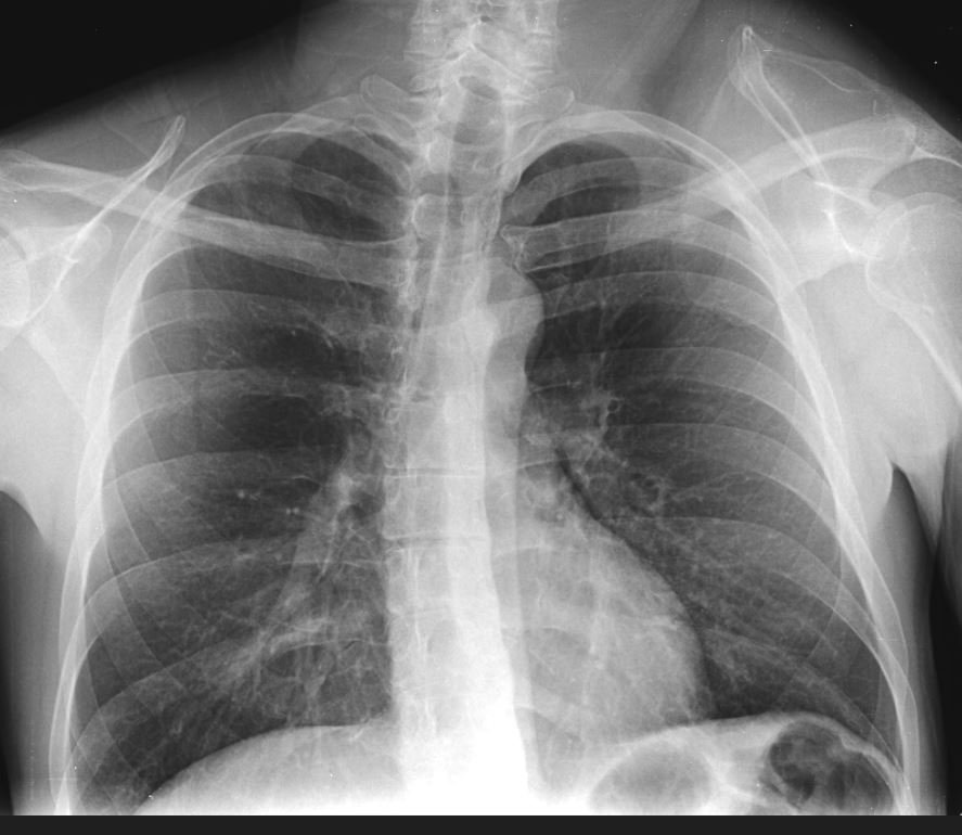
A 43 y.o. man comes to the ED complaining of chest pain.
What do you notice on his CXR? Hint: he has a history of a motorcycle accident.
Our patient had a history of a scapulothoracic dislocation. On the cxr you will notice that the L scapula is superiorly displaced. This event usually occurs with high velocity trauma.
The scapula is fixed to the thorax by muscles, and two joints : the acromioclavicular and sternoclavicular. These can be torn in high speed motor cycle accidents when the rider is attempting to hold on to handlebars while being thrown off resulting in the scapula being disconnected from the thorax.
While our patient had a scapulothoracic dislocation more severe injury can result in a scapulothoracic dissociation where the brachial plexus is torn and vessels are compromised. These patients usually present with massive swelling of the shoulder, a pulseless arm and/or multiple fractures. A shoulder dislocation can be present.
Scapulothoracic dislocation, caused by a less severe injury, can be either extrathoracic as in our patient or intrathoracic with anterior displacement of the inferior angle of the scapula into a post traumatic chest wall defect. There is no shoulder dislocation in this injury. The picture below shows a scapulothoracic dissociation without neurovascularly compromise managed conservatively.
no shoulder dislocation is seen
TO REVIEW
STEPS IN READING A CXR
1. Look for symmetry; it can tell you if there is volume loss, missing ribs, infiltrates or a displaced scapula.
2. Look at the xray as if there are three lungs. Include the trachea as a third “lung” . Lesions in the trachea may be visible as below.
3. Look carefully in the “I” of the cxr; across the clavicles, down the spine and over the diaphragm. Lesions hide easily in these areas.
4. Beware of “ satisfaction of search”. If you find one abnormality keep looking. It is easy to stop looking when you find one abnormality.
The interesting thing about the injury is that muscle spasm will cause the scapula to dislocate again long after the injury. He was managed by rotating the scapula back into place. His chest pain was found to be musculoskeletal.
Kani K, Chew F. Scapulothoracic dissociation. Br J Radiol. 2019 Sep;92(1101) PMC6732935
Hon C, O’Hara C, Little V. endotracheal hamartoma case report: two contrasting clinical presentations of a rare entity. International Journal of Surgery Case Reports. July 2017vol 38. Doi 10.1016/ijscr.2017.07.023
Alharbi A, Abbas A, Mousa W, et al. Scapulothoracic dissociation without a neurologic compromise: a case report. Egyptian Journal of Hospital Medicine July 2022 ;88:3786-3789.
Oreck S, Burgess A, Levine A. Traumatic lateral displacement of the scapula: a radiographic sign of neurovascular disruption. 1984. J Bone Joint Surg Am. 66:758-763.


