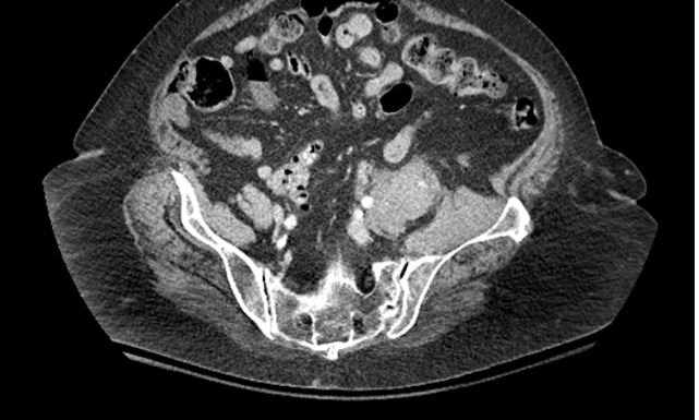
An 80 y.o. woman with a fib, presented with acute back pain radiating down the L leg
No fractures were noted on CT; what do you see?
Our patient had a spontaneous retroperitoneal bleed. While these bleeds usually occur in patients on anticoagulation as in our patient; they can also occur in patients without anticoagulation. In these cases it is thought to be caused by arteriosclerosis of microvessels or vascular lesions such as segmental arterial mediolysis. Segmental arterial mediolysis (SAM) is a nonatheroclerotic arteriopathy characterized by dissecting aneurysms resulting from lysis of the outer media of the arterial wall.
A 9mm pseudoaneursym of the common hepatic artery cased by segmental arterial mediolysis. This is a risk factor for retroperitoneal bleeding.
In patients on anticoagulation, a retroperitoneal bleeding rate of 0.6-6% has been documented with a mortality rate of 20%. Most patients respond to conservative measures with 25% requiring angiography and 9% needing surgery.
the sciatic nerve is the longest nerve in the body and its sensory branches supply all but the medial leg.
While spontaneous retroperitoneal bleeding is relatively uncommon, sciatica is very common with 40% of people experiencing it sometime in their lifetime.. It refers to pain, weakness or tingling in the leg due to injury or pressure on the sciatic nerve. The most common cause is a herniated disc although it can also be caused by spinal stenosis or piriformis syndrome. The pain begins in the buttock where five nerve roots combine to form the sciatic nerve: two from the lumbar spine and three from the sacrum. It radiates down to the foot.
On physical exam, the straight leg raise is often positive. In this test the hip is flexed with the knee extended. The test is positive if pain is reproduced below 60 degrees of hip flexion. As the nerve becomes more damaged there may be weakness in plantar flexion or loss of ankle-jerk reflexes.
Imaging is often negative but can be falsely positive as well. 20-36% of patients who do not have symptoms will have findings on CT.
the sciatic nerve can run through the piriformis muscle causing leg pain in 12-17% of the population
Risk factors for sciatica include: direct trauma to the sciatic nerve, a job requiring prolonged standing or bending forward, heavy manual labor and exposure to vibrations. .
Initial management is 6-8 weeks of conservative treatment unless “red flags” are present. These would include evidence of infection or malignancy.
CLINICAL PEARL
NUMBNESS IN THE BUTTOCKS CAN ALSO BE A SIGN OF CAUDA EQUINA SYNDROME IF IT IS ASSOCIATED WITH BOWEL OR BLADDER DYSFUNCTION.
In our patient, a CT revealed retroperitoneal bleeding. She was managed conservatively and recovered.
Backgaard J, Eskesen T, Lee J, et al. Spontaneous retroperitoneal and rectus sheath hemorrhage-management, risk factors and outcomes. Word J Surg 2019 Aug;43(8):1890-1897.
Chan Y, Morales J, Reidy J. et al. Management of spontaneous and iatrogenic retroperitoneal haemorrhage: conservative management, endovascular intervention or open surgery? In J Clin Pract 2007;62(10):1604-13.
Phillips C, Lepor H. Spontaneous retroperitoneal hemorrhage caused by segmental arterial mdiolysis. Rev Urol. 2006;8(1):36-40.
Miller K. Physical assessment of lower extremity radiculopathy and sciatica. J Chiropr Med. 2007 Spring;6(2):75-82.


