A 35 y.o. Asian male presents with gradual alteration of mental status and inability to speak over 8 weeks
His MRI is shown below.

Hint: the location of the thalami are shown below.

The patient has increased signal in both thalami; What could be the cause?
Although our patient was thought at first to have Wernicke’s encephalopathy, he never improved with high dose thiamine. His diagnosis was changed to limbic encephalitis.
Limbic encephalitis is characterized by inflammation of the brain caused by an autoimmune process. The main regions of the brain involved are the hippocampus, affecting memory, and the amygdala, causing emotional outbursts. Although it is called limbic encephalitis, on autopsy studies the inflammation involves other areas of the brain.
The majority of cases are associated with a tumor. Tumors of the lung, thymus, breast, testes, and ovary may be associated with limbic encephalitis. These receptor antibodies include:
Anti-Hu, associated with small cell carcinoma of the lung
Anti-Ma, associated with germ-cell tumors of the testis
Anti-NMDAR, associated with tumors of the ovaries, often teratomas
Non neoplastic causes include herpes simplex type 1 , AMPA, and GABA receptor antibodies., and voltage-gated potassium channel antibodies (VGKC-Abs). These antibodies react with proteins in the neural cell membranes. The treatment for non neoplastic encephalitis is immune suppression.
Our patient followed simple commands, rarely spoke, had left esotropia, and developed 2nd and 3rd degree heart block requiring a pacemaker. He also developed intractable hiccups and bulbar paralysis requiring intubation. His LP came back positive for SSA/SSB antibodies and was ANA positive confirming autoimmune encephalitis. He is currently on steroids and plasmapheresis was begun.

A 13 y.o. with uptake in the left temporal area from limbic encephalitis.
Molloy A, Cassidy E, Ryan A, O’Toole O. 2011, VGKC positive autoimmune encephalopathy mimicking dementia. BMJ Case Rep. doi:10.1136/bcr08.2011.4642.
Ganesan , Beri S, Khan B, Hussain N. Voltage gated potassium channel antibodies positive autoimmune encephalopathy in a child: A case report and literature review of an under-recognized condition. 2013. Ann Indian Acad Neurol. Oct-Dec;16(4):593-596.
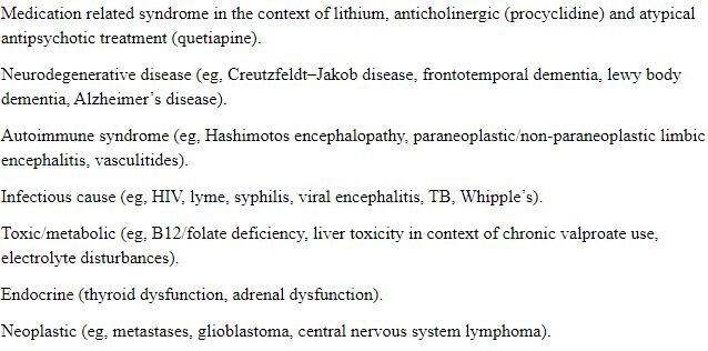
DIFFERENTIAL FOR ENCEPHALITIS.