A 45 y.o. male with CLL presented with a cranial nerve III palsy, diplopia and ptosis
A representative example of his MRI is shown.
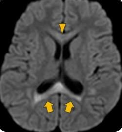
what is his diagnosis?
Our patient had a cytoxic lesion of the corpus callosum (CLOCC). This is a variant of posterior reversible encephalopathy syndrome. (PRES) The lesions are primarily caused by an inflammatory cascade in the brain. This causes a build-up of fluid inside cells (cytotoxic edema) in a highly sensitive area of the brain, the corpus callosum. The corpus callosum contains a dense concentration of receptors for excitatory amino acids like glutamate, and cytokines. It is not a stroke caused by lack of blood flow.
Two things can help differentiate this from a stroke:
1. It is reversible and found in the corpus callosum.
2. It is associated with lethargy and altered mental status that is reversible.
This finding does not reveal the patient’s diagnosis; just that there has been an inflammatory cascade of events that caused an increase in glutamate causing an inflex of water and calcium into neurons and resultant swelling in the corpus callosum. The MRI finding of CLOCC is seen in many things that cause delirium including:
1. Infection- COVID, influenza
2. Seizures -especially with changes in medication
3. Metabolic disturbances-hypoglycemia, high ammonia levels
4. Drugs- clozapine, chemo agents, alcohol withdrawal
5. Trauma- subarachnoid hemorrhage
6. Malignancy- brain tumors
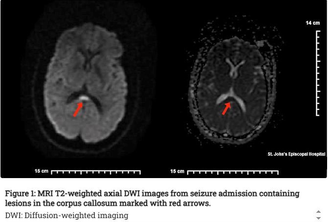
corpus callosal lesion with a seizure
In order to narrow the possible diagnoses in our patient, a broad net was cast. The patient was febrile with neurologic symptoms. He was positive for Ehrlichia which is known to cause an overwhelming inflammatory response in some cases with toxic shock as well as meningoencephalitis.
Ehrlichia is a type of bacterial disease spread primarily through the bite of infected ticks although it can be spread through transfusions and organ transplants as well. In Missouri, the lone star tick is most likely to transmit Ehrlichia.
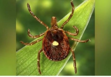
the female lone star tick has a white spot
CLINICALLY
Typically, patients with Ehrlichia complain of flu-like symptoms, fever and headache. A rash, common in other rickettsial diseases like RMSF , is absent in 70% of adults. Thrombocytopenia, low WBC, increased liver functions and occasionally renal failure or meningoencephalitis can develop.
TREATMENT
The first-line treatment for all rickettsial disease is doxycycline. In CLL, infections can be a sign of worsening disease requiring treatment since CLL caused immunocompromise. .
. FUN FACT
Ehrlichia was named in honor of a German scientist Paul Ehrlich who discovered the organism in 1889 in WBCs of guinea pigs. He did not realize they were rickettsial organisms. Howard Ricketts discovered the cause and transmission of rickettsial disease; RMSF in 1906 and typhus in 1909. Ricketts died of typhus while investigating an outbreak.
McCauley C, Li V, Kobrin S. Late-onset seizure disorder in adult cerebral palsy associated with covid-19 infection. Cureus 15(4):e37438
Moors S, Nakhostin D, Lichenko D, et al. Cytotoxic lesions of the corpus callosum: a systematic review. Eur Radiol 2023 Dec26;34(7):4628-4637.
Broppel G, Gallmetzer P, Prayer D, et al. Focal lesions in the splenium of the corpus callosum in patients with epilepsy. Epilepsia 2009 Jun;50(6):1354-60.
Snowden J, Simonsen K. Ehrlichiosis StatPearls Treasure Island FL 2026.
Nichols Heitman K, Dahlgren FS, Drexler NA, Massung RF, Behravesh CB. Increasing Incidence of Ehrlichiosis in the United States: A Summary of National Surveillance of Ehrlichia chaffeensis and Ehrlichia ewingii Infections in the United States, 2008-2012. Am J Trop Med Hyg. 2016 Jan;94(1):52-60