A 73 y.o. male is referred from an OSH for a red eye with decreased vision
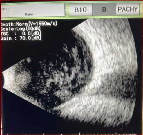
The US of the eye is shown below
What is wrong with his eye?
Our patient had endophthalmitis which is an infection of intraocular fluids.
Most commonly this is due to an exogenous infection: post surgery, after keratitis or a corneal ulcer. In some cases it is endogenous spread hematogenously from an infection somewhere else in the body as in the case of our patient. In the case of endogenous infection: 85% are gram positive bacteria with fungal infections occurring only 5% of the time.
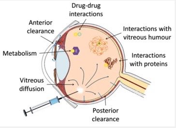
Since the vitreous of the eye has a poor blood supply parenteral antibiotics are not recommended( unless treating a systemic source like a UTI or liver abscess). Intravitreal antibiotics are the treatment of choice. Vitrectomy is only recommended if the vision is “light perception”
intravitreal injections are generally given in the lower outer quadrant of the eye(away from the nose)
To calculate the dose of antibiotics needed the volume of the vitreous is estimated at 4ml. 1,000 micrograms of vancomycin provides a concentration of 250 micrograms/ml.
While not always present, a hypopyon is a clue that endophthalmitis is present. This is a layering of white cells in the anterior chamber of the eye. It is not specific and can be present in eye trauma, uveitis, and autoimmune diseases like Behcet’s.
a hypopyon
Our patient grew MSSA in the blood and vitreous fluid. He was given intravitreal injections of vancomycin and ceftazidime. He was given Oxacillin q 4 hours after cultures resulted and no source was found for his bacteremia. TEE was negative for vegetations and no evidence of UTI or liver abscess was found. He is scheduled to receive Oxacillin q 4 hours for six weeks.
Rodriguez A, Gonzalez B, Teijeito B. A review of intraocular pharmacokinetics of anti-infectives commonly used in the treatment of infectious endophthalmitis. Pharmaceutics 2018 10(2)
Simakurthy S, Triperthy K, Endophthalmitis. Treasure Island , FL2024 Jan 2023 Aug 25.
Durand M. Endophthalmitis. Cinical Microbiology and Infection Vol 19(3)March 2013;227-234.


