A 74 y.o male with nonischemic cardiomyopathy, a fib and renal failure present s after CRT D therapy with nausea and epigastric pain
CRT D involves trying to synchronize the atrium and ventricle with a pacemaker consisting of three leads: the R atrium, R ventricle and coronary sinus. The “D” part includes a defibrillator if the EF is < 30.

this is the patient’s cxr
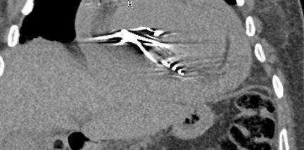
what do you notice on the noncon CT?
Our patient has a large pericardial effusion with perforation of the RV by the pacer wire.
Our patient underwent CRT D (resychronization therapy with a defibrillator) This is recommended in patients with symptomatic heart failure and L ventricular EF of < 35% in spite of maximal medical management. This is also known as biventricular pacing and involves simultaneous pacing of the RV and LV with a coronary sinus lead placed for LV pacing. The procedure was introduced in the 1990s and multiple randomized controlled trial show that it improves heart failure symptoms. The QRS represents ventricular depolarization and a prolonged QRS(>120 ms) is a marker of ventricular dyssynchrony. The net result is decreased pump function when the ventricles fire over a longer time. While CRT is recommended for LBBB, it has not been shown to be useful in RBBB.

wither a pacemaker alone or a pacer/defibrillator can be placed. CRT—D describes the defibrillator
Our patient developed elevated lactate and acidosis with hypotension. He underwent emergency pericardiocentesis.
How common is it to perforate the RV with a pacemaker lead?
The incidence of post pacemaker implantation perforation of the RV is 0.3-1.2% which goes up to 5% if an AICD is placed. The perforation usually manifests in 24 hours but multiple reports of delayed perforation exist. While perforation of the RV can cause hemopericardium as in our patient; in 15% of cases there is no pericardial bleeding or tamponade. Other symptoms have been reported such as hiccups or chest wall contractions as the diaphragm or chest wall may be paced.

sometimes the pacer wire perforates without tamponade
Our patient became hypotensive and one liter of blood was drained from the pericardium. His blood pressure rose to 120-140. He was given K centra and had a drain placed . Gradually the bleeding slowed. He has been discharged and will not be anticoagulated for several weeks.
Yamamoto A, Takahashi S. Delayed right ventricular lead perforation by a pacemaker lead 2-year post implantation. Clin Case Rep 2022 Apr;10(4)e05760
Udo E, Zuithoff N,van Hemel N. Incidence and predictors of short and long-term complications in pacemaker therapy: the FOLLOWPACE study Heart Rhythm. 2012;9:728-735.
McAlister F, Ezekowitz J, Hooton N, et al. Cardiac resynchronization therapy for patients with left ventricular systolic dysfunction:a systematic review. JAMA 2007 Jun 13 297(22)::2502-14.
Glikson M, Nielsen J, Kronborg M, et al. for the ESC Scientific Document Group. 2021 ESC. Guidelines on cardiac pacing and cardiac resynchronization therapy Europace. 2021 Aug 29.