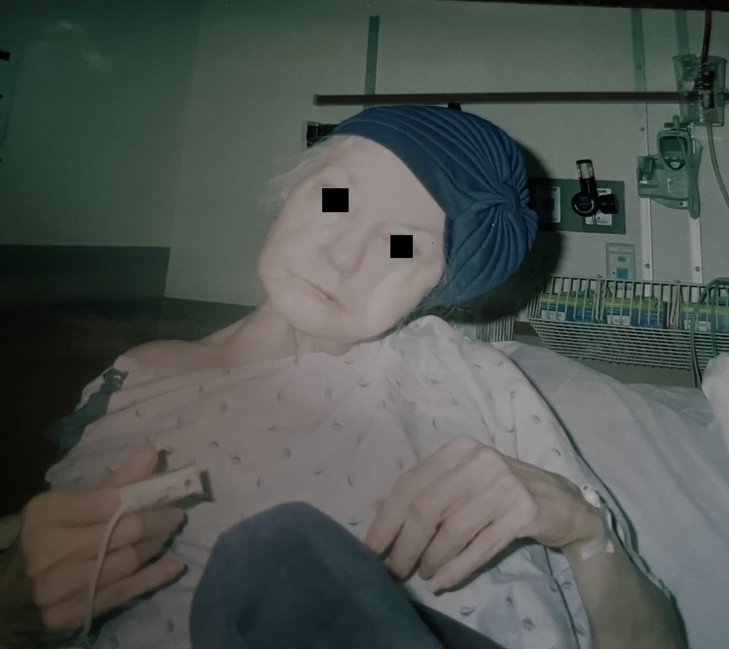
A 76 y.o. woman comes in with a chief complaint of "my head fell off"
what happened to her?
the patient is unable to move her head to the R
Our patient had an atlantoaxial dislocation. This results when the atlas (C1)and the axis (C2) lose their stable articulation. This can happen from trauma, inflammatory and idiopathic causes. It occurs in all age groups but is most often seen in adolescents.
Since over 60% of cervical spine rotation occurs at the dens; great flexibility is provided in this area. C1 and C2 are the only vertebral bodies that do not bear the weight of the skull. The load is transferred to the lateral masses of C1 and then on to the lateral masses of C2. These are nearly horizontal articulations and depend on ligaments to provide stability. When the ligaments are weakened, the head can fall to the side.
The left shows normal alignment of the dens and C1; the R shows ligamentous laxity.
In addition to traumatic injuries; there are conditions that predispose to atlantoaxial dislocation. In Down’s syndrome,juvenile rheumatoid arthritis and ankylosing spondylitis the ligaments supporting the dens are weakened. Basilar invagination, where the dens protrudes into the foramen magnum (as in Chiari malformation), Infections and retropharyngeal abscesses can also cause atlantoaxial dislocation. Subluxation of the C1-C2 joint has been described with cervical and tonsillar abscesses. It was first reported in 1830 and caused by syphilitic ulceration of the pharynx. This is referred to as Grisel’s syndrome.
Grisel’s syndrome is acquired ligamentous laxity following an infection.
An 11-year-old girl with a fixed torticollis and right hand weakness on a background of >1 month of neck pain and recurrent tonsillitis
While atlanto axial dislocation (AAD) requires immediate neurosurgical consultation; a similar appearing condition, torticollis, does not.
Torticollis presents with the head rotated to one side. It is the result of tightening of the neck muscles and may be present at birth or develop in adulthood as a result of trauma, medication, dystonia or structural changes in the spine. In the case of muscular torticollis; often relief of muscle spasm will correct the rotation.
In order to distinguish between the two conditions AAD or torticollis; first determine the history and on physical. First, note which side the spasm is on. In rotatory dislocation, the sternomastoid spasm is on the side to which the chin twists where in muscular torticollis the spasm is on the opposite side. This is thought to be because the spasm with a dislocation is the muscle trying to compensate for being stretched to the side of the dislocation. While xray can be difficult to interpret; CT will demonstrate a dislocation.
Our patient had atlantoaxial dislocation because of rheumatoid arthritis. Her chin was rotated L with spasm on the L side. She was unable to move her head to the R. She underwent a similar surgical procedure to the one pictured below.
The ligamentous laxity is now fixed.
Greenberg M, Forgeon J, Kurth L. et al. Atlantoaxial rotatory subluxation presenting as acute torticollis after mild trauma. Radiology Case Reports 2020 Vol 15(11) :2112-2115.
Yang S, boniello A, Poorman C, et al. A review of the diagnosis and treatment of atlantoaxial dislocations. Golbal Spine J, 2014 Aug;4(3):197-210
Kuroki H, Kubo S, Hamanaka H, et al. Posterior occipito=axial fixation applied C laminar screws for pediatric atlantoaxial instability caused by Down syndrome: report of 2 cases. Int J of Spine Surgery. 2-12 Vol 6210-215.
Goel A. Torticollis and rotatory atlantoaxial dislocation : a clinical review. J Craniovertebr Junction Spine 2019 Apr-Jun;10(2)::77-87.
C. Bell The nervous system of the human body: embracing the papers delivered to the Royal Society on the subject of the nerves Longman, Rees, Orme, Brown, and Green, London (1830)


