A 62 y.o. male with a hx of previous resection of a benign brain tumor comes to the ED with headache and fever.
what do you notice on his CT?
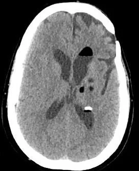
Our patient had a ruptured dermoid cyst with a lipid/fluid interface in the ventricle. Dermoid cysts are present at birth and occur when ectoderm layers don’t grow together normally. They contain remnants of ectodermal tissue including sebaceous cysts, oil producing hair follicles, squamous epithelium and sometimes teeth. They most commonly present in the first three decades of life. Dermoids are the most common pediatric tumor and are found in the orbit and periorbital areas often over the L eyebrow.

the lipid/csf interface from a ruptured dermoic
Dermoids are benign tumors but as they grow they can compress surrounding structures. They can occur In any area of the body. In the ovary, as the size of the ovary increases torsion may occur. They are often resected but grow back at a rate of 3-4%.
In the brain dermoids account for <1% of all intracranial masses They are most commonly found in the midline because they are felt to be ectopic ectoderm from the neural tube. They are often in the suprasellar cistern. As they grow they are at risk for rupture and serious complications such as chemical meningitis, vasospasm and infarct can occur.
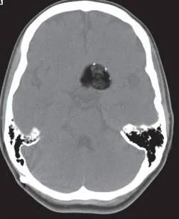
An unruptured dermoid presents as a fatty lesion in the brain.
RANDOM FACT
Dermoids can be thought of as tumors along a spectrum with epidermoid cysts at one end ;containing only squamous epithelium and teratomas at the other; containing elements of all three embryonic layers. Dermoid cysts are true hamartomas which are defined as an abnormal mixture of mature cells which are found in a region where they normally occur.
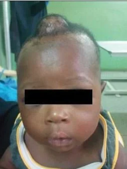
Dermoids can be found where the skin fails to fuse normally as at the anterior fontanelle.
Our patient is still hospitalized. He had a ventricular drains placed for progressive hydrocephalus, aseptic meningitis was diagnosed, and hyponatremia developed. This was treated with tolvaptan. He is awake and alert with slight expressive aphasia with drains still in place.
Pollard Z, Harley R, Calhoun J. Dermoid cysts in children. Pediatrics. 1976Mar;57(3):379-82.
Jackow J, Tse G, Martin A, et al. Ruptured intracranial dermoid cysts: a pictorial review. Pol J Radiol 2018;83:e465-e470. Doi: 10.5114/pjr 2018.80206.
MucajS, Ururel M, Dedushi K, et al. Role of MRI in diagnosis of ruptured intracranial dermoid cyst, case report. Acta Inform Med. 2017;25:141-144.
Adenigba P, Lawal T, Elemile P, et al. The value of ultrasonography in the diagnosis of a rare congenital dermoid cyst of the anterior fontanelle in an infant. Journal of the West African College of surgeons 2019. Vol 9(4):21-25.