A 43 y.o. male presents with eye pain
What do you notice on CT images of the orbits?
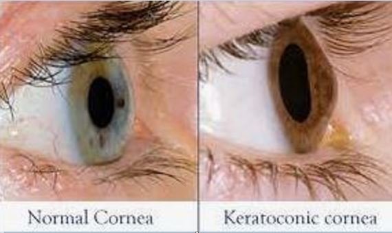
Our patient had keratoconus on imaging with bulging of the cornea. the normal round shape of the globe is lost.
Keratoconus is a weakening of the collagen of Descemet’s membrane in the cornea leading to thinning of the cornea and scarring. This leads to irregular astigmatism, central scarring and reduced vision. It occurs in approximately one in 2,000 individuals beginning in puberty.
Layers of the cornea
In some cases hydrops of the cornea develops. A break in the Descemet membrane occurs and aqueous humor leaks into the stroma and cause stromal edema. A white patch of stromal edema is then visible on the corneal. 3% of people with hydrops have corneal perforation and it is important to diagnose.
A white patch appears on the cornea in keratoconus with hydrops.
Scleral lens are used to treat keratoconus. Scleral lenses were initially made out of glass until poly-methyl methacrylate was available in the 1940’s which put the user at risk for corneal hypoxia. Gas permeable scleral contacts became available in the 1980’s. The scleral lens differs from a regular contact lens in that there is a fluid reservoir under the lens which fills in any defects in the cornea and thereby improves vision.
In the case of hydrops, the standard treatment previously was topical treatment or surgery followed by a scleral lens. Recently, gas has been injected into the anterior chamber blocking the extrusion of aqueous humor into the stroma anteriorly. This allows the corneal to heal sooner.
Elevated intraocular pressure is a risk factor for keratoconus and our patient had both glaucoma and keratoconus. The pt was seen several more times for glaucoma and eye redness with vision loss in spite of being on four classes of drugs for his glaucoma. He missed his appointment in eye clinic.
Harthan J, Shorter E, Therapeutic uses of scleral contact lenses for ocular surface disease: patient selection and special considerations. Clin Optom (Auckl) 2018;10:65-74.
Gialousakis J, Management of acute corneal hydrops in a patient with keratoconus: a teaching case report. Optometic Education: Volume 45 (2) Winter-Spring 2020.
Romero-Jimenez, Santodomingo-Robido, et al. Keratonconus: a review. Cont Lens Anterior Eye. 2010 Aug: 33(4):157-66.
Maharana P, Sharma N, Vajpayee R. Acute corneal hydrops in keratonconus. Indian J Ophthalmol 2013;61(8):461-4.
McMonnies C. Mechanisms for acute corneal hydrops and perforation. Eye Contact Lens. 2014;40(4):257-264.


