A 2 y.o. with a history of Medium-chain acyl-CoA dehydrogenase deficiency presents with neck pain after falling from the bottom bunk
How would you read his CT?

The patient refused to move his neck because of pain.
Our patient had an epidural hematoma which was unrecognized on the first visit. Ten days later he was having difficulty moving his arms and his mother brought him back. Review of the first CT at that time showed an epidural hematoma which had been missed.

While there are four types of spinal hematomas: epidural, subdural , subarachnoid and intramedullary, by far the epidural hematoma is most common because of the rich vascular supply of the epidural space. There are no valves in the epidural veins and this makes them more likely to rupture with changes in pressure. 40% of epidural hematomas are idiopathic presenting clinically as pain or weakness, so a history of trauma is not needed to consider the diagnosis.

While an epidural abscess can be seen on CT as in the case of our patient, the imaging modality of choice is the MRI.
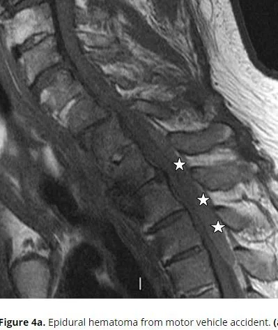
MRI however, changes over time as hemoglobin changes its magnetic properties in various stages of evolution. So while in the acute phase blood is hypointense on both T1 and T 2; after ten days it will become hyperintense on both T1 and T2 signals.

Our patient underwent an emergency laminectomy from C3-T1 with evacuation of the epidural hematoma. His arm strength improved and he was discharged with outpatient Pt follow up.
Pierce J, Donahue J, Nacey N, et al. Spinal hematomas: what a radiologist needs to know . Radiographics. 2018 https://doi.org/10.2248/rg.2018180099
Thiel W. Supplement to the conservation of an entire cadaver according to W. Thiel. Ann Anat 2003;184(3): 267-269.
Westbrook J. Anatomy of the epidural space. Anaesth Intensive Care Med. 2012;13(11):551-554.