A 59 y.o. woman was hit in the head with a pipe at work; five days later she was found on the floor at her home
What does the CT show?

why is she weak on the R side if the subdural is on the right?
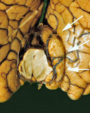
Our patient had a Kenohans’ notch herniation: pressure on the opposite sided cerebral peduncle causing right sided subdural with RIGHT sided weakness. This is a form of uncal herniation and requires immediate neurosurgical attention.

weakness on the same side as the lesion
There are two classic brain herniation syndromes: uncal herniation and infratentorial tonsillar herniation. Since the volume of blood, brain and CSF are constant an increase in blood results in a decrease in volume of brain or csf. The brain is displaced from one brain compartment into another.
Uncal herniation occurs when the uncus (medial temporal lobe) herniates medially into the tentorial notch causing compression on the third nerve and then the brainstem as it progresses. There is a same sided third nerve palsy causing a fixed dilated pupil ( a blown pupil) on the side of the lesion and contralateral hemiparesis. Uncal herniation is treated with placing the head of the bed up at 30 degress, hypertonic saline or mannitol and ultimately decompressive craniectomy. An external ventricular drain is often used.
herniation of the medial temporal lobe causing compression of the brainstem
Tonsillar herniation occurs when the cerebellar tonsils herniate through the foramen magnum. This results in the Cushing reflex consisting of irregular breathing, bradycardia and hypertension. Tonsillar herniation is treated with posterior decompression and often results in death.

cerebullum heriating through the foramen magnum represented by the white line
Our patient underwent decompressive craniectomy and then developed herniation again from an epidural hematoma resulting in a second surgery. She remains lethargic.

types of herniatiion
Plum and Posner described a sequence of progressions of the brain during herniation. ADDITIONAL PEARLS
This is referred to as a rostral-caudal progression.
1. Subfalcine herniation- the cingulate gyrus migrates under the falx anteriorly resulting in infarctions along territory of the anterior cerebral artery.Lower limb weakness occurs.
2. Transsphenoidal herniation-either compression of the middle cerebral artery against the sphenoid or internal carotid artery compression against the anterior clinoid
3. Trans uncal herniation – leads to compression of the third nerve with dilation of the same sided pupil. Sometimes the contralateral cerebral peduncle is forced against the tentorium resulting in paralysis on the same side as the lesion as in our case.
4. Central herniation- ascending and descending herniation occurs as the brainstem continues to descend. Flexor and extensor posturing is noted. Breathing changes from cheyne stokes as the diencephalon is affected to central neurogenic hyperventilation at the medulla is compressed.
5. Cerebellar tonsillar herniation- The cerebellum is forced through the foramen magnum causing ischemia of the brainstem.
Stevens R, Shoykhet M, Cadena R. Emergency Neurological Life Support: Intracranial Hypertension and Herniation. Neurocrit Care. 2015;23 Suppl 2:S76-82.
Plum F, Posner J. The diagnosis of stupor and coma. Contemp Neuro Ser 1972;10:1-286.
Sauvigny T, Gottsche J, Czorlich, et al. Intracranial pressure in patients undergoing decompressive craniectomy: new perspective on threshols. J Neurosurg 2018Mar;128(3):819-827.
Dash H, Chavali S. Management of traumatic brain injury patients. Korean J Anesthesiol 2018 Feb;71(1):12-21.