A 38 y.o. woman with renal failure comes in with L hip pain.
her plain film was read as normal


what do you notice on her CT?
Our patient had two processes going on: iliopsoas tendonitis and lytic lesions called brown tumors.
Iliopsoas tendonitis refers to inflammation of the tendon which acts as a flexor of the hip. It drapes over the front of the hip socket and if it is inflamed it can cause a painful “click” over the lesser trochanter. This occurs commonly in athletes, dancers, in 4% of people having hip surgery and , as in our patient, in patients with bone disease. This was confirmed in our patient with a CT scan.

The second thing confirmed on CT scan was the presence of lytic lesions of the femur and pelvis.
Our patient was on dialysis and had renal osteodystrophy. Renal failure causes excessive Ca excretion which lowers serum Ca and increases parathyroid hormone. This stimulates osteoclasts and causes bone loss with areas of bleeding and fibrosis. These areas appear as lytic lesions on Xray and are called “brown tumors” because of hemosiderin deposition.
THE DIFFERENTIAL FOR LYTIC LESIONS ON XRAY
Metastases/multiple myeloma/primary bone tumors
Bone infarct(osteomyelitis or tuberculosis)
Eosinophilic granuloma
Fibromas
Giant cell tumors.
Sarcoid
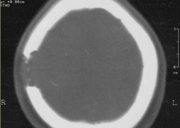
eosinophilic granuloma causing a lytic lesion in the skull
Our patient was also found to have a 5 cm mass in the R breast which she had known about for two years. She declined further imaging.
Howarth D, Gilchrist G, Mullan B. et al. Langerhans cell histiocytosis: diagnosis, natural history, management and outcomes. 1999 Cancer May 15; 85(10) 2278-80
Di Lorenzo L, Jennifer Y, Pappagallo M, Psoas impingement syndrome in hip osteoarthritis. Joint Bone Spine. 2009;Jan;76(1):98-100.
Anderson C. Iliopsoas: Pathology, Diagnosis, treatment. Clin Sports Med 2016 Jul;35(3):419-433.
Reynolds D, Hernang Y, Utz J. 57y.o man with hip pain and lytic bone lesions. 2016 Mayo Clinic Proceedings 91(3)3e35-e40.