A 39 y.o. woman with lupus presents with swollen breasts and no other swelling
What could be wrong?
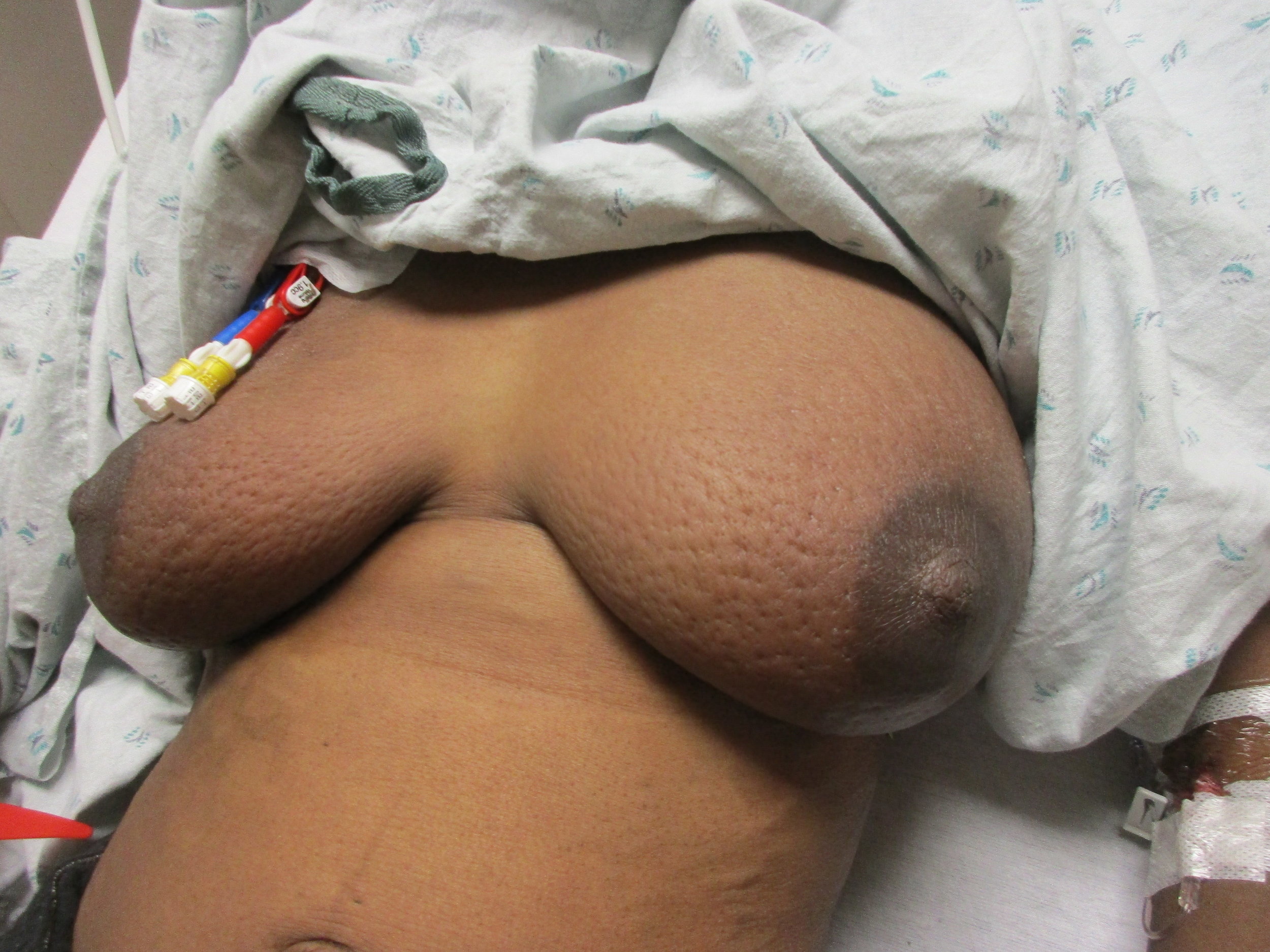
Our patient had classic peau d’orange, but had superior vena caval syndrome and not inflammatory breast cancer. She did not have swelling of her face because of extensive chest wall collaterals.
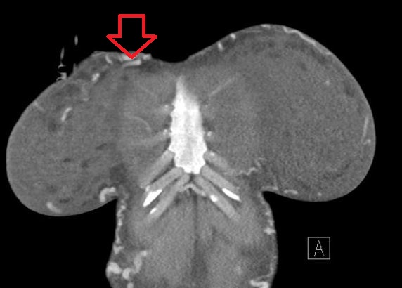
collaterals on the chest wall with svc thrombosis.
It was first described in 1757 by William Hunter in a patient with syphilitic aortitis compressing the SVC. The syndrome occurs in 15,000 people in the US a year. The most common cause is malignancy with 85% reported to be caused by malignancy in 2008 but now 40% are reported in non malignant cases due to the prevalence of ports. Other causes of SVC syndrome include: fibrosing mediastinitis, tuberculosis, histoplasmosis , actinomycosis and thrombosis caused by pacemaker leads.

fibrosing mediastinitis causing svc obstruction
Shaikh I, Berg K, Kman N. Thrombogenic Catherter-Associated Superior vena cava syndrome. 2013, Case Resports in Emergency Medicine Volume 2013, article ID 79054, 3 pages.
Higdon M, Higdon J. The treatment of oncologic emergencies. Am Fam Physician 2006;74:1873-80.
Wilson L, Dettebach F, Yahalom. Superior vena caval syndrome. 2007 NEJM 356:1862-1869.
Cohen R, Mena D, Carbajal-Mendoza A, et al. Superior vena caval syndrome: A medical emergency? Int J. Angio 2008 spring 17(1):43-46