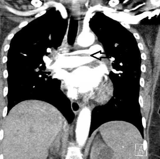
A 61 y.o. female has had a COPD exacerbation x several weeks
She was watching the superbowl, using crack cocaine and heroin and on returning home developed lightheadedness and tingling on the L side of her body. She passed out on her lawn when arriving home and was incontinent of stool. A TPA page was called on arrival in the ED. Head CT was negative.
What do you notice on her chest CT?
Our patient had a saddle embolus. In addition to her strange presentation, more history was obtained . She had a myelodysplastic disorder and was hypercoagulable. She had a previous PE and was supposed to be on Coumadin which she had not taken in over a week. She also had a clot while therapeutic on Coumadin so an IVC filter was placed on this admission.
The patient had persistent L sided weakness and tingling and although her MRI was neg she was felt to have a stroke. How common is a stroke with negative diffusion weighted MRI? An “invisible stroke?”
There has been a significant increase is the use of MRI in the diagnosis of stroke. It was thought at one time to be 88-100% sensitive for diagnosing stroke but recent evidence shows diffusion weighted MRI fails to identify stroke in 30% of cases. The specific areas where it fails are in three categories: the posterior fossa, the brainstem, and in hyperacute ischemia( within six hours of symptoms). These patients with negative imaging have the same outcomes as strokes with positive imaging; persistent deficits and risk of recurrent strokes.
The reason for “invisible strokes” may be that the reduction in cerebral blood flow required to initiate cell swelling( and a positive MRI) is more severe than that required to produce neurologic symptoms.
Case thanks to Dr. Ruoff, with stroke information supplied by Dr. Panagos.


