A 43 y.o. woman with previous back pain comes in with R leg pain.
What is the circular finding on her MRI at L4-5 ? she has no weakness or sensory loss.
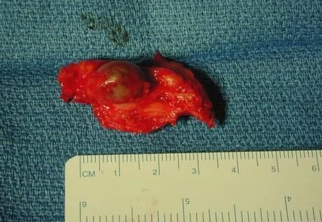
Our patient had a synovial cyst. Once again Dr. Docherty is correct) A synovial cyst in the spine is a fluid filled sac resulting from degeneration of a facet joint. They were first described in 1885 by Baker( who also described the one behind the knee). On MRI they appear well demarcated and are extradural. They are more common in women. The cysts are most often found at the L4,5 level because that is the area of maximum mobility. They grow very slowly and often are asymptomatic but they can cause radicular symptoms, spinal stenosis or even cauda equina.
synovial cyst
Surgery is indicated for intractable symptoms. The complications of surgery include cerebrospinal fluid fistula, discitis, epidural hematoma, seroma, deep vein thrombosis and death. Our patient is scheduled for surgery because of intractable pain.
Baker’s cyst behind the knee
Khan A, Girardi F. spinal lumbar synovial cysts. Diagnosis and management challenge. Eur Spine J v 15(8) 2006:1176-1182.
Baker W. formation of synovial cysts in connection with joints. St. Batholomews Hospital Reports. 1885;21:177-190.
Baker W. On the formation of synovial cysts in the leg in connection with disease of the knee-joint. 1887. Clin Orthop. 1994;299:2-10.
Lyons M, et al. Surgical evaluation and management of lumbar synovial cysts: the Mayo Clinic experience. J Neurosurg. 2000;93.1(supple):53-57.


