A 40 y.o. male with a history of aortic dissection repair and paraplegia comes to the ED with altered mental status.
an abnormality was noted on cxr.


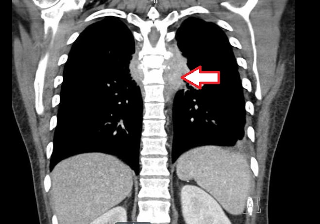
And confirmed on CT. What is the problem?
Our patient had a spinal epidural abscess which unfortunately had not been treated for some time.
He had a history in the past of an MVC in which he sustained an aortic injury and became paraplegic. He presented from a long term care facility with altered mental status and a widened mediastinum was noted on CXR. The CT showed the epidural abscess. In spite of antibiotics, he died of sepsis.
Epidural abscess is often not recognized on the first ED presentation. The “red flags” for the diagnosis include: unexplained fever, focal neurologic deficit , immunosuppression, IV drug use, prolonged steroid use, weight loss, enduring back pain and history of cancer. In 58% of the cases with errors, the “red flag” was documented but not acted upon.. The route of infection was identified in 52% of caseswith bacteremia as the most common cause (26%) followed by recent surgery(21%), and spinal injection. (6%). The most common risk factors were steroid use and diabetes. The most common organism was Staph Aureus, with MRSA in 25%. . The incidence is 5 cases for every 10,000 admissions in a study by Vakili however, our ED has seen at least three cases in the past month.
The second case (shown above) was a schizophrenic; well known for ingesting foreign objects and stabbing himself in the abdomen. He presented with a fever and paraplegia. His abscess wrapped around the aorta and esophagus and he was deemed “inoperable” by thoracic surgery. He remains paralyzed and in the hospital two months after presentation awaiting placement.

A third was a woman with diabetes who had previous back surgerywhich became infected one month pta. She presented to an outside hospital with nausea and vomiting and CT showed a psoas abscess. She was noted to have acute cholecystitis which was drained percutaneously and was discharged on long term IV antibiotics. Candida tropicalis grew from the gall bladder drain and nothing grew from the psoas abscess ( she had been previous treated with antibiotics.)
Bhise V et al. Errors in diagnosis of spinal epidural abscesses in the era of electronic health records. Am J Med 2017 Aug; 130:975.
Vakili M, Crum-Cianflone N. Spinal Epidural Abscess: A series of 101 Cases. American Journal of Medicine 2017, in press.
Davis DR, Wold RM, Patel RJ et al. The clinical presentation and impact of diagnostic delays on emergency department pateitns with spinal epidural abscess. J Emerg Med 2004;26(#):285-291.
Darouich RO. Spina epidual abscess. N engl J Med. 2006;355:2012-2020.