A young woman with fever, generalized weakness and a new rash....
A young woman with Crohn's disease is brought in by ambulance for one day of fever and malaise. On arrival, she is febrile to 39.1˚C, tachycardic, hypotensive and tachypneic. She appears acutely ill. Chest X-ray and urine are clear. Her abdomen is non-tender, but given her clinical history and presentation, a CT scan of the abdomen is obtained to evaluate for a source of infection.

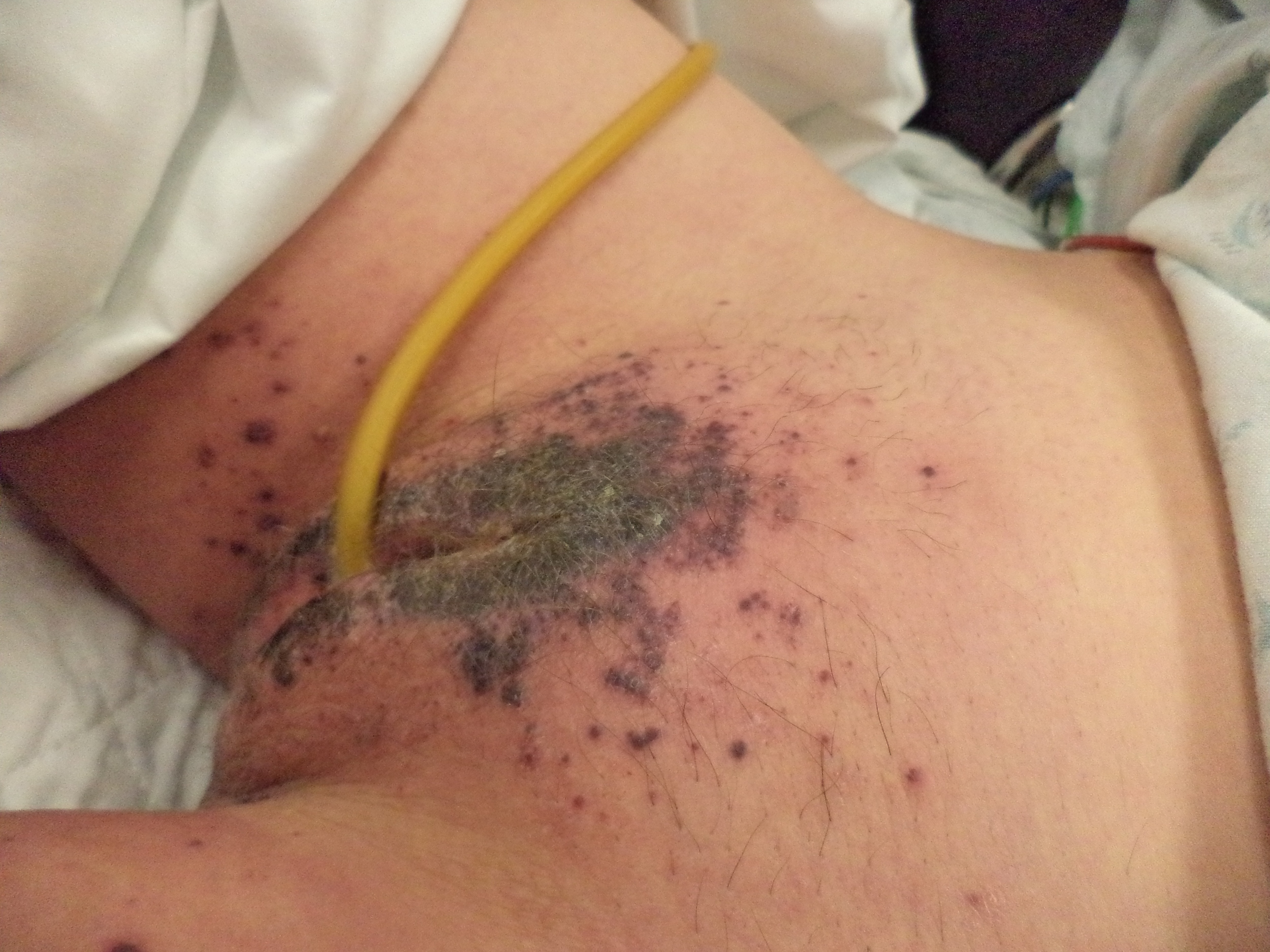
She is aggressively resuscitated with IVF and broad spectrum antibiotics. During this resuscitation, her nurse notes that she is developing a rash around her perineum and buttocks.
What is your differential diagnosis for this patient? What tests would you send and what would you do next?
Scroll Down for the Case Conclusion.
Final Diagnosis: Purpura Fulminans due to Streptococcus pneumoniae bacteremia
Case Conclusion: The patient was treated with broad spectrum antibiotics and aggressively resuscitated. Lab work revealed thrombocytopenia, low fibrinogen, coagulopathy, and elevated d-dimer consistent with disseminated intravascular coagulation (DIC). The patient went into mult-system organ failure requiring ventilatory support, dialysis and vasopressors. Blood cultures grew Strep pneumoniae. It was determined that the patient's major risk factor for this infection was congenital asplenia (see CT scan above), and she was vaccinated against Neissieria and Pneumococcus after recovery and prior to discharge from the hospital.
Learning Points: Purpura Fulminans(PF) is the result of an acute inflammatory response early in sepsis and is most commonly seen in pediatric or younger patients [1]. In sepsis, it is a clinical manifestation of DIC in which widespread activation of the coagulation pathway and consumption of anticoagulant proteins (especially Protein C) leads to widespread microvascular thrombosis. Microvascular thrombosis in the skin is indicative of a systemic process, and affected patients are likely to also have hemorrhagic infarction in other organs, especially the lungs, kidneys, central nervous system and adrenal glands. The development of PF is therefore a harbinger of impending multi-system organ failure. Mortality rate in patients with PF exceeds 40% [2].

Protein C deficiency is the final common pathway of the multiple etiologies of Purpura Fulminans, including congenital, infectious, autoimmune, and drug-induced causes .
The initial clinical appearance of PF is well-demarcated, erythematous macules that progress rapidly to irregular areas of purple-black hemorrhagic necrosis surrounded by a thin border of erythema. The differential diagnosis for these initial lesions also includes thrombotic thrombocytopenia purpura (TTP), immune-complex vasculitis (such as Henoch-Schonlein Purpura), ehrlichiosis and other rickettsial infections [3]. As patients begin to bleed into a necrotic dermis, these lesions become raised and more painful. Eventually, this process can lead to full thickness skin or soft tissue necrosis [1].
Asplenia is a major risk factor for PF because of the increased risk of developing overwhelming sepsis with encapsulated organisms. This is because splenic macrophages phagocytose circulating bacteria and splenic lymphocytes are a major producer of antibodies [4]. Congenital asplenia usually occurs in the context of another genetic syndrome, but isolated congenital asplenia has been reported in multiple case reports of patients presenting with overwhelming sepsis [5].
The most important treatment of PF is to treat the underlying cause, including rapid initiation of broad-spectrum antibiotics and circulatory support. In addition to IV fluids and vasopressors, patients may need stress dose steroids given the risk of adrenal infarction. FFP should be used to replete consumed coagulation and anti-coagulation factors, particularly Protein C and S [1,2]. When PF is accompanied by large vessel venous thrombosis, heparin may be administering cautiously and concurrently with FFP [1].
Case Conclusion by Maia Dorsett (@maiadorsett), PGY-4
References:
1.Chalmers, E., Cooper, P., Forman, K., Grimley, C., Khair, K., Minford, A., ... & Mumford, A. D. (2011). Purpura fulminans: recognition, diagnosis and management. Archives of disease in childhood, 96(11), 1066-1071.
2. Wojtowicz, J. M., & Jones, G. L. (2014). Streptococcus pneumoniae–induced purpura fulminans in a woman with functional asplenia. CJEM, 16(04), 339-342.
3. Thorner, A. R., Walker, D. H., & Petri Jr, W. A. (1998). Rocky Mountain spotted fever. Clinical infectious diseases, 1353-1359.
4. Brigden, M. L. (2001). Detection, education and management of the asplenic or hyposplenic patient. American family physician, 63(3), 499-506.
5. Gilbert, B., Menetrey, C., Belin, V., Brosset, P., de Lumley, L., & Fisher, A. (2002). Familial isolated congenital asplenia: a rare, frequently hereditary dominant condition, often detected too late as a cause of overwhelming pneumococcal sepsis. Report of a new case and review of 31 others. European journal of pediatrics, 161(7), 368-372.